Introduction
Hemangiosarcoma is an aggressive and often fatal cancer that affects dogs. It arises from the cells that line blood vessels in the body and can spread rapidly through the bloodstream to other organs. While hemangiosarcoma only accounts for 5-7% of all cancers in dogs, it is unfortunately one of the deadliest. The average survival time after diagnosis is only about 6-8 months if treated with surgery alone.
However, early detection of hemangiosarcoma can significantly improve outcomes. When caught early while still localized, surgical removal of the tumor can prolong survival to 12 months or more. This highlights the importance of owners being vigilant for any signs of illness in their dogs and discussing diagnostic testing with their veterinarian. Bloodwork is one component of screening for hemangiosarcoma, but has limitations in detecting this cancer in its early stages.
What is Hemangiosarcoma?
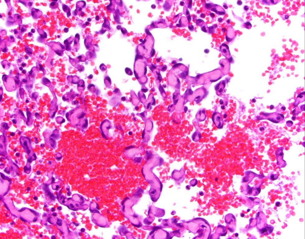
Hemangiosarcoma is an aggressive and malignant cancer of blood vessels that originates from the cells lining the blood vessels, known as endothelial cells (1). It is one of the most common cancers diagnosed in dogs, primarily affecting the spleen, heart, and liver (2). Hemangiosarcoma can spread quickly through the body as the cancerous endothelial cells have direct access to the bloodstream. This makes it very difficult to detect early and contributes to its aggressive nature with poor prognosis.
The spleen is the most common organ affected by hemangiosarcoma in dogs. The spleen contains an extensive network of blood vessels which provide an optimal environment for this cancer to grow. Hemangiosarcoma in the spleen may not cause any obvious clinical signs initially. Heart-based hemangiosarcoma in dogs can lead to pericardial effusion or cardiac tamponade which can be life threatening. Hemangiosarcoma tumors in the liver can also grow quite large before causing any noticeable symptoms.
Overall, hemangiosarcoma is known for being difficult to detect in the early stages. Dogs can deteriorate rapidly once significant clinical signs develop, as the cancer has often spread or impacted major organs by that point.
Importance of Early Detection
Catching hemangiosarcoma early has been shown to greatly improve prognosis and survival times for dogs. According to research, the median survival time for dogs diagnosed with stage 1 localized hemangiosarcoma and treated with surgery alone is around 6-8 months, whereas median survival drops to only around 2-3 months if allowed to progress untreated to stage 2 metastasis [1]. The longer survival times associated with early intervention highlight the critical importance of early detection and swift action.
Early detection also opens up more treatment options that can further lengthen survival. If caught early while still localized, surgery can remove the primary tumor and provide temporary control. But once metastasis occurs, surgery is no longer an option. Chemotherapy has also shown improved efficacy when administered to dogs with stage 1 disease rather than later stage [2]. New investigational treatments being developed, like immunotherapy vaccines, rely on early detection as well in order to initiate therapy while the immune system is still strong enough to mount a response [3].
Overall, initiating any form of treatment or intervention early on provides the best chance of slowing disease progression, managing symptoms, and extending a dog’s life. Hemangiosarcoma can appear and spread rapidly, so prompt diagnosis and action as soon as the first subtle signs appear gives owners and veterinarians the most options for maximizing their dog’s remaining time and quality of life.
Sources:
[1] https://www.akc.org/expert-advice/health/hemangiosarcoma-in-dogs/
[2] https://www.ncbi.nlm.nih.gov/pmc/articles/PMC10093745/
[3] https://www.ethosdiscovery.org/blog-post/hemangiosarcoma-developing-new-treatment-options/
Limitations of Bloodwork
Bloodwork alone often fails to detect hemangiosarcoma in the early stages. While abnormalities may eventually appear as the tumors grow and spread, they are often not present when the cancer first develops. As noted in a study published in Preprints, “These limitations emphasize the potential value in the ability to detect [hemangiosarcoma] prior to the emergence of clinical signs or clinicopathologic abnormalities.”[1] So while bloodwork is a useful diagnostic tool, it has significant limitations for identifying hemangiosarcoma in its initial stages.

The tumors can become quite large before bloodwork reveals any issues. According to the NIH, hemangiosarcoma tumors are often at an advanced stage by the time they are detected.[2] This underscores the need for additional testing beyond standard bloodwork to screen for this aggressive cancer early on.
Additional Diagnostic Tests
Since hemangiosarcoma develops from the endothelial cells that line blood vessels, bloodwork alone cannot definitively diagnose hemangiosarcoma. Additional diagnostic testing is necessary to confirm a diagnosis. Standard imaging tests like x-rays, ultrasound, and MRI can help identify tumors and lesions associated with hemangiosarcoma [1]. However, the gold standard for diagnosis is examination of tissue samples obtained via biopsy.

Ultrasound is a useful initial imaging test for detecting hemangiosarcoma tumors in the spleen, liver, and heart. Ultrasound can identify the location and characteristics of tumors, which appear as hypoechoic nodules or masses [2]. However, ultrasound alone cannot definitively diagnose hemangiosarcoma.
MRI provides high resolution soft tissue imaging to visualize tumors in locations like the heart and central nervous system. MRI has high sensitivity for detecting splenic masses and can also assess for metastasis in the liver and peritoneal cavity [1].
To confirm diagnosis, biopsy samples of the tumor must be taken for microscopic examination. This allows a veterinary pathologist to analyze the tumor cells and architecture to definitively identify hemangiosarcoma. Needle aspiration and surgical biopsies are standard methods for obtaining samples [3]. However, biopsies of highly vascular tumors like hemangiosarcoma carry some risk of bleeding complications.
Bloodwork as Part of Diagnosis
While bloodwork alone cannot definitively diagnose hemangiosarcoma in dogs, it does provide part of the overall diagnostic picture when combined with other tests. Bloodwork allows veterinarians to look for non-specific abnormalities that, together with additional diagnostics, can point towards hemangiosarcoma. For example, bloodwork may reveal anemia, low protein levels, or high liver enzymes – changes that should prompt further investigation into potential causes like cancer.1
Tracking bloodwork over time is also useful for monitoring the progression of hemangiosarcoma and evaluating the effectiveness of treatment options. Specific blood markers can provide insight into whether the cancer is spreading or responding to therapy. So while blood tests alone cannot confirm a hemangiosarcoma diagnosis, they remain an important component of the diagnostic workup and ongoing management when combined with medical imaging, cytology, biopsy, and other veterinary assessments.
Bloodwork Details
There are several blood tests that may indicate the presence of hemangiosarcoma in dogs:
- Complete blood count (CBC) – May show low red blood cell count (anemia), low platelet count (thrombocytopenia), or high white blood cell count.
- Biochemical profile – Elevated liver enzymes may indicate metastasis to the liver.
- Coagulation tests – Prolonged clotting times can indicate bleeding disorders associated with hemangiosarcoma.
However, many dogs with hemangiosarcoma have normal blood work initially. Over time, bloodwork may become abnormal as the cancer progresses and causes internal bleeding, anemia, clotting issues, etc. So while blood tests can provide clues, normal results do not rule out hemangiosarcoma.
Additional diagnostic imaging such as x-rays, ultrasound, or CT scans are often needed to directly visualize tumors and detect hemangiosarcoma.
Treatment Options
Treatment for hemangiosarcoma typically involves a combination of surgery, chemotherapy, and radiation:
Surgery
When possible, surgical removal of the tumor is recommended. This can prolong survival times significantly if the cancer has not already spread. Surgery aims to fully remove the primary tumor and any affected lymph nodes (CVM NCSU).
Chemotherapy
Chemotherapy is often used after surgery to target any remaining cancer cells. The most commonly used chemotherapy drug for hemangiosarcoma is doxorubicin. Studies show chemotherapy can extend survival times by up to 6 months on average (De Nardi et al., 2023).
Radiation
Radiation therapy may be used in combination with surgery and chemotherapy. It is most helpful for localized tumors that have not metastasized.
Palliative Care
For advanced stage or metastatic hemangiosarcoma that cannot be cured, the goal is to control symptoms and improve quality of life. This may involve medications for pain relief, anemia, blood clots, etc. Providing comfort care is essential.
Prognosis
Unfortunately, even with treatment, the prognosis for dogs diagnosed with hemangiosarcoma is poor. Hemangiosarcoma is an aggressive cancer that spreads rapidly and most often affects the liver, spleen, heart and skin. The median survival time for dogs after diagnosis depends on a few factors.
For dogs with splenic HSA that undergo splenectomy alone, the median survival time is only 1-3 months[1]. With splenectomy followed by chemotherapy, median survival time improves to 6-8 months[2]. Dogs with cardiac HSA treated with surgery and chemotherapy have a median survival time of 4-6 months. Subcutaneous HSA has a more favorable prognosis with a median survival time of around 1 year with surgery.
While these median survival times may seem short, it’s important to remember that some dogs can live much longer than these estimates with treatment, care and a bit of luck. But unfortunately the aggressive nature of this cancer means most dogs will succumb within 6-12 months of diagnosis.
Conclusion
In summary, while bloodwork alone is insufficient to definitively diagnose hemangiosarcoma in dogs, it can provide clues that may prompt further testing when combined with a veterinary exam and imaging tests. Some of the key points to remember are:
- Hemangiosarcoma is an aggressive blood vessel cancer often found in the spleen, liver, heart or skin of dogs.
- Early detection is critical, but challenging due to the hidden and rapid spread of tumors.
- Bloodwork may show non-specific abnormalities like anemia, thrombocytopenia or an inflammatory response.
- Additional diagnostic tests like ultrasounds, x-rays, CT scans and biopsy are required for definitive diagnosis.
- A multi-modal approach with bloodwork, imaging and biopsy is recommended for the highest chance of early diagnosis.
- Treatment options include surgery, chemotherapy and supportive care to extend quality of life.
While the prognosis for dogs with hemangiosarcoma remains guarded, the best outcomes rely on veterinary teams using all available tools for early detection and swift intervention. Bloodwork can provide warning signs to trigger further action, and remains a useful part of the diagnostic process when combined with other tests.