Introduction
X-rays are an important diagnostic tool used by veterinarians to examine a dog’s internal body structures. They allow vets to visualize the internal anatomy and detect potential problems or abnormalities that may not be apparent from a physical exam alone. X-rays are commonly used to diagnose bone fractures, arthritis, intestinal obstructions, tumors, pneumonia, and other conditions. They provide valuable information to vets about a dog’s skeletal structure, organs, and tissues. X-rays are non-invasive and one of the most common diagnostic imaging techniques used in veterinary medicine today.
This article will provide an overview of the reasons vets use x-rays for canine patients, the safety precautions taken, the x-ray procedure from start to finish, and how vets analyze the images to reach a diagnosis. Understanding the process and importance of x-rays will help dog owners appreciate this key tool that aids veterinarians in promoting canine health.
Reasons Vets Use X-Rays
One of the most common reasons vets use x-rays is to diagnose injuries or illness in dogs. X-rays allow vets to see inside a dog’s body and evaluate the bones, joints, organs, and soft tissues. They are especially helpful for identifying broken bones, arthritis, intestinal obstructions, tumors, pneumonia, and other conditions that may not be obvious from a physical exam alone.
For example, if a dog has been limping or crying in pain after an injury, x-rays can identify if there is a broken bone or joint damage. The images give the vet crucial information to determine the best treatment approach. X-rays are also used to monitor healing and ensure bones are aligning properly after fractures.
When a dog is vomiting, has diarrhea, or lacks appetite, abdominal x-rays can pinpoint potential causes like swallowed foreign objects, tumors, ulcers, or intestinal blockages. Without imaging, these issues can be difficult to diagnose. X-rays provide vital insight so vets can address the underlying problem.
In cases of respiratory distress like coughing or shortness of breath, chest x-rays allow vets to visualize the lungs, heart, and major blood vessels. They can identify conditions like pneumonia, fluid buildup, heart disease, and cancer for proper treatment.
Overall, x-rays are an essential tool for vets to accurately diagnose a wide variety of orthopedic, gastrointestinal, respiratory, and other internal issues in dogs that may not be apparent upon examination. They provide crucial insight into a dog’s health and allow for tailored treatment plans.
Safety Precautions
Taking X-rays involves exposing dogs to radiation, so vets take precautions to limit radiation exposure. According to South Wilton Veterinary Group, the amount of radiation used in a typical X-ray is very small. However, vets still aim to limit radiation exposure as much as possible.
Vets use lead aprons and thyroid shields to protect dogs’ bodies from radiation exposure. They also collimate the X-ray beam so only the area being imaged is exposed. The veterinary staff leaves the room during the X-ray to avoid radiation exposure. Proper shielding and positioning helps focus the radiation only on the area of interest.
According to Fairway Knolls Veterinary Care, X-rays account for a small fraction of most pets’ total radiation exposure. But vets still take every precaution to minimize radiation from veterinary X-rays.
The X-Ray Room
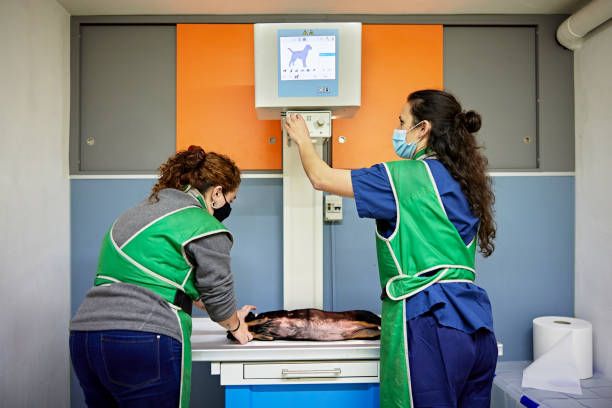
The x-ray room at a veterinary clinic is specially designed to safely take x-rays of animals. It contains an x-ray machine, an x-ray table, and protective shielding on the walls, ceiling, windows and door (https://www.xraycurtains.com/blogs/news/shielding-your-veterinary-office-during-x-ray-procedures). The shielding is typically lead lined and prevents radiation from escaping the room, protecting staff and other animals in the clinic.
The x-ray machine is on wheels so it can be easily maneuvered around the table. The table where the animal lays is also adjustable, allowing the technician to position the dog properly for the x-rays. There may be sandbags, foam wedges, and other positioning aids to help hold the dog still.
There will be monitors and computer equipment in the x-ray room to capture the digital images and allow the technician to see the finished x-rays. The computers also contain software to process and manipulate the x-ray images as needed.
Overall, the x-ray room is designed to be safe for taking diagnostic images of animals while protecting staff from repeated radiation exposure.
Positioning the Dog
Properly positioning the dog is crucial for getting clear and useful x-ray images. The positioning will vary depending on the area being x-rayed.
For x-rays of the head, the dog should be placed in dorsal recumbency with the front legs pulled caudally and secured with tape. The dog’s nose should be centered on the table and the beam should be centered on the head area of interest (Source).
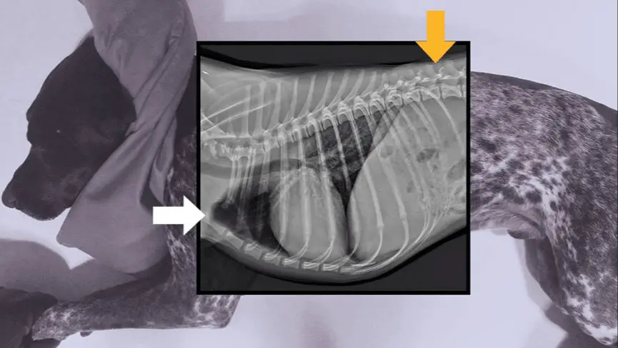
For thoracic x-rays, the dog should be placed in right lateral recumbency with the legs pulled forward. The beam should be centered vertically on the chest. Forelimb x-rays require the dog to be in lateral recumbency with the relevant leg pulled forward and up. The beam should be centered over the area of the leg being examined (Source).
Abdominal x-rays need the dog in dorsal recumbency, ideally with the rear legs pulled caudally. The beam needs to be centered vertically and horizontally on the abdomen. Proper positioning is essential for visualizing all the abdominal organs clearly.
For the hind limbs, the dog should be placed in lateral recumbency with the relevant hind leg pulled forward and down, keeping the stifle and hock at 90 degree angles. The beam should focus directly on the area of the leg being x-rayed.
Sedation
Sedation may be necessary to keep the dog calm and still in order to get clear x-rays. According to https://mgqi.throwbackcity.org/, sedation is sometimes required in order to obtain quality images that the vet can accurately interpret. Lively, anxious, fearful, or active dogs are often sedated so they remain motionless during the x-ray process. This prevents motion blur and allows the x-ray equipment to capture sharp images of the skeletal structure and organs.
Puppies also frequently require sedation because they tend to squirm and resist staying still. Small dogs can be difficult to position without sedation too. For the safety and comfort of the dog, vets may decide that sedation leads to the best possible x-ray results.
Taking the X-Rays
Taking x-rays of a dog is a quick and straightforward process that the veterinarian will guide you through step-by-step. Here is what you can expect during this part of the appointment:
First, the vet will bring your dog into the x-ray room and gently position them on the table in the needed poses. They may lightly restrain your dog to ensure they hold still long enough to get a clear shot. Your vet will put on a lead apron to protect themselves from repeated radiation exposure.
For most x-rays, sedation is not required. The vet will calmly speak to your dog and give them treats to keep them relaxed. However, if your dog is extremely anxious, sedation may be recommended.
Once your dog is steady, the vet will step behind a protective wall or window, set the x-ray machine, and take the exposure. They will take at least two views (from the side and from the front or back) to get comprehensive images. The x-ray only takes a split second.
Your dog will need to change positions for each view. The vet will gently reposition them and repeat the process until all needed images are captured. Taking x-rays is quick, with the full set typically completed within 10 minutes. The number of x-rays depends on the area being examined.
Digital radiology systems allow vets to see the x-rays immediately and retake any poor-quality images right away. With traditional film, the x-rays must be developed before they can be examined, which takes additional time.
Throughout the brief x-ray process, the vet will reassure your dog, reward them with treats, and make it as stress-free as possible. With the vet’s guidance, taking x-rays is easy for dogs!
Developing the Images
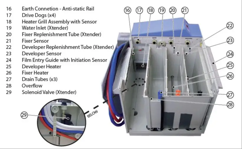
After the X-ray images are taken, the films need to be developed in order to view the internal structures of the dog. The X-ray films are placed into a machine called an automatic processor which develops the images. This machine contains chemicals such as developer solution and fixer solution which process the films.
The process involves the following steps:
- The undeveloped films enter the processor machine and move through a series of chambers containing developer solution. This brings out the latent image on the film.
- The films then pass through a chamber containing water to wash off the developer solution.
- Next, the films move through a chamber containing fixer solution which sets the image permanently on the film.
- Another wash cycle removes the fixer solution.
- Finally, the films pass through a drying cabinet before exiting the processor fully developed.
The entire process from start to finish usually takes around 90 seconds. The automatic film processor allows the veterinary staff to quickly develop the X-ray films in-house so they can be analyzed right away by the veterinarian.
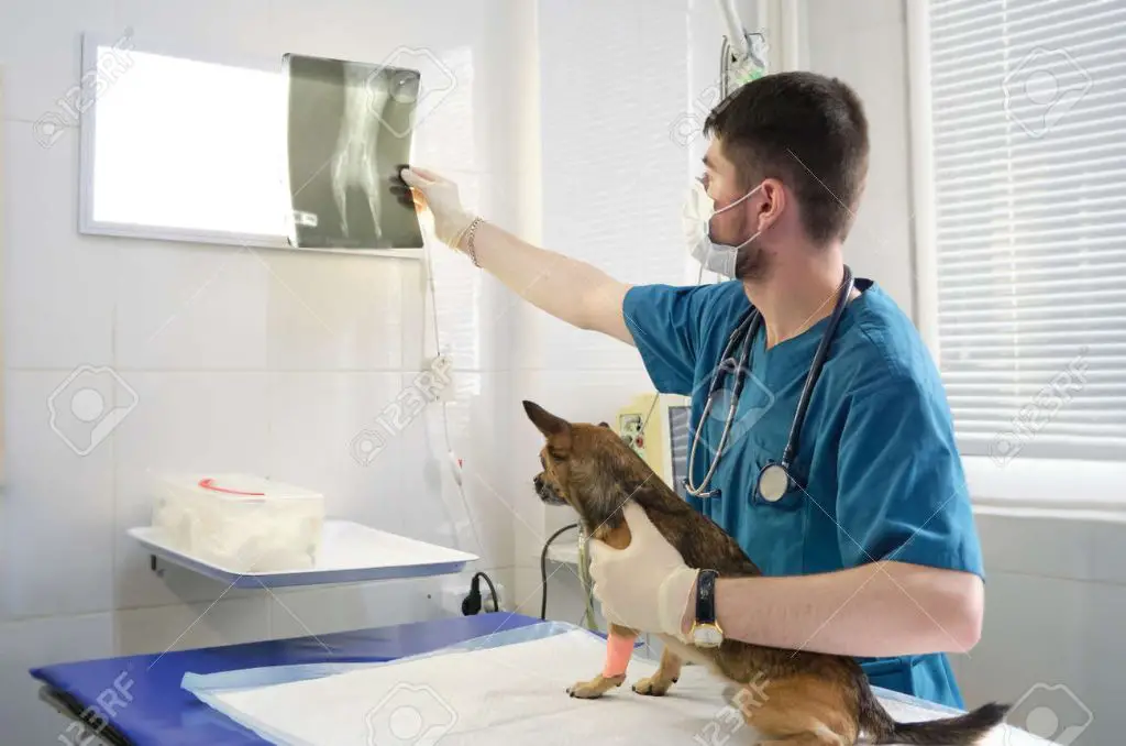
Reading the X-Rays
Once the X-ray images have been developed, the veterinarian will carefully analyze them to diagnose any underlying issues. Reading and interpreting X-rays is a skill that vets develop with years of training and practice. They will look for any areas of concern like breaks, fractures, foreign objects, tumors, etc. The Essential Role Of X-Rays In Veterinary Care For Dogs And Cats explains that vets must consider things like the dog’s age, breed, and medical history when examining X-rays. For example, certain breeds are prone to specific joint issues that a vet would look for. The anatomy of dogs can vary greatly between breeds as well, so the vet must have extensive knowledge of canine physiology. If the images are not clear or the vet needs a different view, additional X-rays may be taken until the issue is identified. With experience reading dog X-rays, vets can quickly diagnose many common conditions like arthritis, fractures, swallowed foreign objects, and more.
Conclusion
X-rays provide important diagnostic information for vets to assess a dog’s health and detect issues such as broken bones, tumors, and swallowed foreign objects. While x-rays involve a cost, they are a key tool vets rely on to gain insight into a dog’s internal health and structure. The process involves careful positioning and sedation when necessary to capture high-quality images to inform treatment. Reading x-rays takes training and an experienced eye. With appropriate safety measures, x-rays are an invaluable, non-invasive procedure with the power to potentially save dogs’ lives through early detection and diagnosis.