Introduction
X-rays are a critical diagnostic tool used by veterinarians to detect issues and abnormalities in dogs that are otherwise undetectable from physical exams alone. By passing x-rays through a dog’s body, veterinarians can create photographic images that reveal the dog’s skeletal structure as well as organs and tissues. These internal views allow vets to diagnose bone fractures or dislocations, tumors, fluid buildup, foreign objects, and signs of many diseases. X-rays are painless procedures that require minimal restraint and provide valuable information to inform treatment plans. They help veterinarians evaluate a dog’s overall health and wellbeing.
Bone Issues
X-rays are commonly used to diagnose various bone disorders and conditions in dogs. Fractures, arthritis, and bone cancer are some of the main bone issues that can be identified on x-rays.
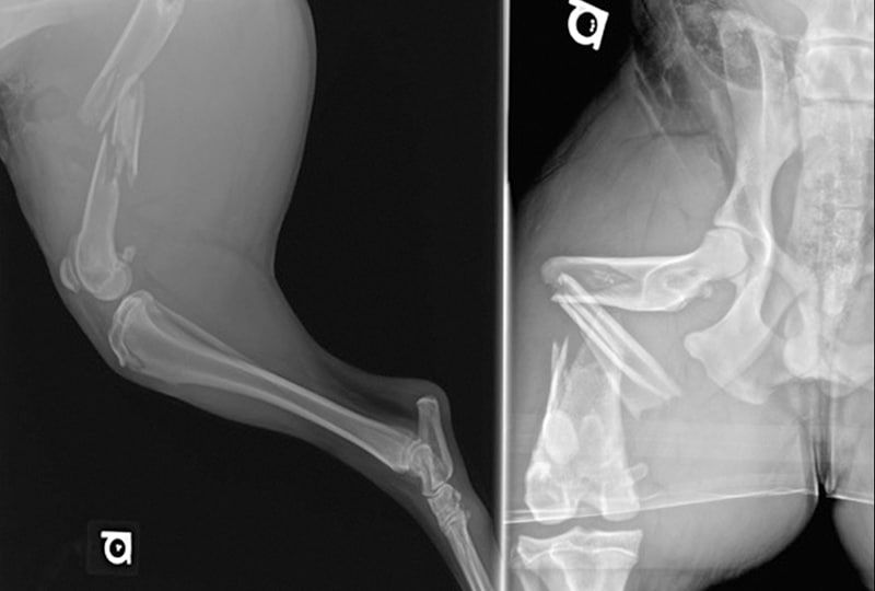
Fractures show up on x-rays as a disruption in the normal smooth contour of the bone. The type and location of the fracture provides information for determining the best treatment approach. Fractures may be described as transverse (straight across the bone), oblique (on an angle), spiral, or comminuted (bone broken into multiple pieces). X-rays allow veterinarians to assess the degree of displacement and alignment of the bone fragments (Seiler).
Osteoarthritis, also known as degenerative joint disease, is characterized by deterioration of the cartilage in joints. On x-rays, vets look for signs like new bone formation, cartilage loss, and changes to the joint space. The joints most commonly affected are the hips, elbows, and knees. X-rays help determine the severity of arthritis and guide treatment options like medication, joint supplements, weight loss, or surgery (Merck Veterinary Manual).
Primary bone cancer and cancer that has metastasized to bone can also be detected on x-rays. Signs include bone destruction, cortical bone disruption, and proliferation of abnormal tissue. Common types of bone cancer seen in dogs include osteosarcoma, chondrosarcoma, and hemangiosarcoma. Early detection via x-ray gives the best chance for successful treatment (MSPCA).
Organ Issues
X-rays can help diagnose various issues with a dog’s organs, including enlarged organs, tumors, and obstructions. One of the key organs vets look at is the heart. X-rays allow vets to see the size and shape of the heart and identify conditions like cardiomegaly (an enlarged heart), which could be a sign of heart disease.
X-rays can also reveal masses or tumors in organs like the lungs, liver, spleen, and kidneys. Lung tumors may show up as abnormal masses, while tumors in other organs can cause the organ to appear enlarged on x-ray. X-rays can help locate tumors and determine their size and spread.
For the gastrointestinal system, x-rays are useful for finding foreign objects, obstructions, and blockages. Things like bones, rocks, socks, or other inedible items that a dog swallowed may be visible on x-ray. X-rays can also show intestinal obstructions that prevent proper digestion.
So in summary, when it comes to organ issues, vets rely on x-rays to diagnose enlarged organs, masses, tumors, and obstructions affecting the heart, lungs, digestive system, and other vital organs.
Joint Issues
X-rays can help diagnose various joint issues in dogs such as dysplasia, injuries, and arthritis. Hip dysplasia is one of the most common joint diseases in dogs and x-rays are used to evaluate the fit of the femoral head into the hip socket to look for signs of subluxation and osteoarthritis (https://www.ncbi.nlm.nih.gov/pmc/articles/PMC7152260/). Elbow dysplasia is also commonly diagnosed via x-rays by looking for osteophytes around the joint and narrowing of the joint space (https://www.vin.com/doc/?id=8249676).
X-rays allow veterinarians to diagnose fractures, luxations, and other traumatic injuries to joints and the surrounding bones. Signs like irregular bone margins, disruption of joint spaces, and misalignment of bones are clear indicators of injury on x-rays.
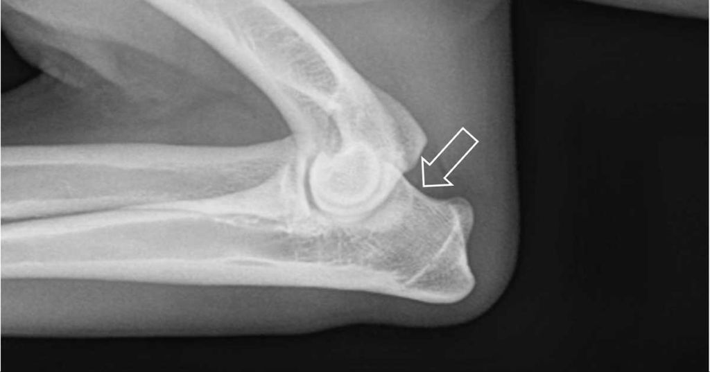
One of the earliest signs of arthritis visible on x-rays is new bone formation seen as bony proliferations called osteophytes at joint margins. Joint effusion and reduced joint space are also seen as arthritis progresses (https://www.vettimes.co.uk/article/x-ray-tips-for-joint-disease-in-canine-thoracic-limbs/). X-rays are useful for monitoring the progression of arthritis over time.
Gastrointestinal Issues
X-rays can help diagnose many gastrointestinal issues in dogs, including blockages, foreign objects, tumors, and inflammation. One of the most common uses of x-rays is looking for intestinal obstructions or blockages [1]. Signs of a blockage may include vomiting, diarrhea, and abdominal pain. On x-rays, blockages appear as obstructions in the intestinal tract. Contrast x-rays using barium can provide additional detail on the location and nature of the blockage [2].
X-rays can also detect foreign objects that a dog may have ingested, such as rocks or toys. These appear as radiopaque densities on the films. Surgical removal is often required for intestinal foreign bodies [3].
Abdominal x-rays may also reveal gastrointestinal tumors, typically seen as soft tissue masses or irregular thickening of the intestinal wall. However, additional diagnostic tests like biopsies are usually needed to confirm cancer.
Respiratory Issues
X-rays are commonly used to diagnose respiratory issues in dogs such as pneumonia, lung infections, and fluid accumulation in the lungs (pulmonary edema). Pneumonia shows up on x-rays as areas of increased opacity in the lungs which indicate infection and inflammation. Miliary pneumonia causes a mottled appearance from many small spots in the lungs. X-rays can also reveal fluid accumulation or a collapsed lung. According to the Merck Veterinary Manual, chest x‐rays are typically done for dogs with lower respiratory signs such as cough, rapid shallow breathing, or labored breathing [1].
One common respiratory issue in dogs is pneumonia, which is an inflammation of the lungs usually caused by a bacterial, viral, or fungal infection. Pneumonia will show up on x-rays as cloudy/white areas called infiltrates. The pattern, location, and distribution of these infiltrates can help determine the underlying cause. For example, the bacteria Bordetella causes a bronchopneumonia pattern affecting the bronchi and bronchioles. Diagnostic x-rays are important for determining the severity of pneumonia and monitoring improvement with treatment.
Another respiratory issue that x-rays can detect is pulmonary edema, which is an accumulation of fluid in the lungs. This causes a diffuse interstitial to alveolar lung pattern on x-rays. Pulmonary edema has various causes including left-sided heart failure, lung disease, trauma, or certain toxins. X-rays allow veterinarians to identify and monitor the amount of lung fluid to guide treatment.
Cardiac Issues
X-rays can help diagnose various heart problems in dogs, including an enlarged heart, heart tumors, and fluid buildup around the heart (pericardial effusion) [1]. An enlarged heart, known as cardiomegaly, indicates the heart muscle has thickened abnormally. This is often caused by high blood pressure or diseases affecting heart function. X-rays allow vets to see the shape and size of the heart shadow and compare it to normal values.
Heart tumors are rare but can be visualized on x-rays as distinct masses. Tumors are most commonly found in the right atrium. Pericardial effusion refers to fluid accumulation around the heart, compressing heart function. Radiographs reveal an enlarged, globoid cardiac silhouette when large volumes of pericardial fluid are present.[2] Identifying pericardial effusion early is crucial, as it can progress to life-threatening cardiac tamponade if left untreated.
While x-rays may detect abnormalities, other diagnostic tests like echocardiograms are usually recommended to thoroughly evaluate the heart and determine the underlying cause.[3] Still, radiography serves as an accessible first-line screening tool for potential heart issues in canine patients.
Other Issues
X-rays can help diagnose a variety of other issues and conditions in dogs beyond just bones, organs, joints, and the gastrointestinal and respiratory systems. Two examples of other issues that may be revealed or examined with x-rays include:
Bladder stones
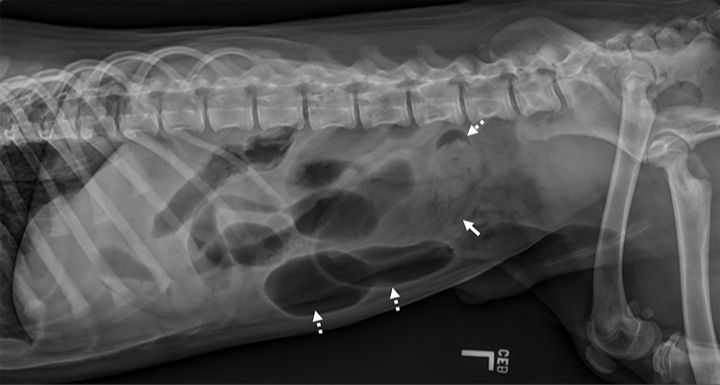
X-rays can reveal the presence of bladder stones (uroliths) in dogs. Bladder stones show up clearly on x-rays as obvious round opacities in the bladder area. Identifying bladder stones is important so they can be properly treated and removed to avoid complications like urinary blockages or a ruptured bladder. According to the VCA, most bladder stones in dogs are radiopaque, meaning they are visible on x-rays. But some stones are radiolucent and may require other imaging tests for diagnosis.
Pregnancy checking
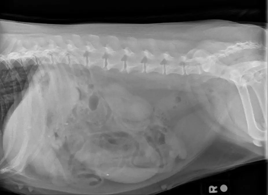
Abdominal x-rays can help confirm pregnancy in dogs by allowing visualization of fetal skeletons in advanced stages of gestation. X-rays are most useful for pregnancy diagnosis in the final 2-3 weeks before delivery when fetal bones have calcified enough to be seen. Earlier in pregnancy, ultrasound is more commonly used to check for fetuses. According to the Carolina Veterinary Specialists, x-rays can provide an estimate of litter size in late pregnancy by counting visible puppy skeletons. However, the number of puppies seen may be less than the total born.
Limitations
While x-rays are a valuable diagnostic tool, they do have some limitations compared to other imaging techniques like CT scans and MRIs:[1]
X-rays are not able to provide the same detailed, high-resolution images that CT and MRI scans can produce. X-rays only visualize bones and organs, while CTs and MRIs can also image soft tissues in great detail. So x-rays may miss certain issues like small tumors or abnormalities in muscles, tendons, or nerves that do not affect the bones or organs directly.
X-rays are also limited to 2D images from a certain orientation. CT and MRI scans allow 3D reconstruction and viewing from any angle. This can help identify issues that may be obscured on a standard x-ray image.
While x-rays are excellent for assessing bone fractures, joint issues, dental problems, and organ enlargement, they cannot match the detail provided by CT and MRI for soft tissue assessment and very small abnormalities. However, x-rays are more readily available, faster, and less expensive than advanced imaging.
Conclusion
In summary, x-rays are an essential diagnostic tool for dogs. They allow veterinarians to see inside a dog’s body in a non-invasive way and identify issues with bones, organs, gastrointestinal system, respiratory system, heart, and joints. X-rays can detect fractures, tumors, foreign objects, pneumonia, heart disease, arthritis, and many other conditions. While x-rays have some limitations, such as not being able to diagnose all soft tissue problems, they provide crucial information to vets and enable targeted treatment plans. The ability to take x-rays has saved many dogs’ lives and improved their quality of life by diagnosing diseases early. When used alongside other diagnostic tools like blood tests and ultrasound, x-rays give vets a comprehensive picture of a dog’s health.