What is the Third Eyelid in Dogs?
The third eyelid, also called the nictitating membrane, is a protective layer of tissue located inside the eye. It sits in the inner corner of a dog’s eye, behind the lower eyelid (Source 1). The purpose of the third eyelid is to help spread tears and lubricate the surface of the eye, as well as protect it from damage (Source 2). It can sweep horizontally across the eye to remove debris and keep the surface moist. Anatomically, the third eyelid originates from the “third eyelid gland” and contains cartilage for support. It provides an extra layer of protection for a dog’s vulnerable eyes.
Normal Third Eyelid Color and Appearance
In healthy dogs, the third eyelid is usually a pale pink or light pink color. It should not protrude from the eye or be visible when the eye is fully open, according to the Washington State University College of Veterinary Medicine [1]. The third eyelid should retract completely into the corner of the eye when the dog blinks.
The third eyelid, also called the nictitating membrane, is a protective layer that lubricates and clears debris from the eye. When healthy, it is located in the inner corner of the eye socket and is normally not visible, according to VetChick [2].
Abnormal Third Eyelid Color Meanings
The color of a dog’s third eyelid can indicate potential health issues. Some key abnormal colors to watch for include:
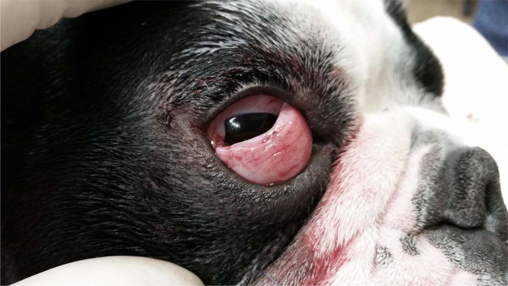
Red/pink: This often signals inflammation or infection of the third eyelid or surrounding structures. Common causes are conjunctivitis, third eyelid gland prolapse (cherry eye), and trauma to the area (Source).
Dark red/brown: This coloring may indicate an ulcer, tumor, or other trauma affecting the third eyelid tissue. Melanomas and other masses can appear dark red or brown (Source).
Black: A black third eyelid is a major red flag for melanoma. Melanomas often originate on the third eyelid before spreading elsewhere (Source).
Yellow: Yellowing or jaundiced hues point to liver disease, jaundice, or other conditions causing an accumulation of bilirubin. This warrants rapid veterinary assessment (Source).
Blue: A bluish tint indicates a lack of oxygen reaching the tissues. This requires emergency veterinary care (Source).
Causes of Abnormal Third Eyelid Color
There are several potential causes of an abnormally colored third eyelid in dogs:
Infection – Infections of the eye or third eyelid can cause redness and inflammation. Common culprits include bacterial infections, viral infections like canine herpesvirus, and parasites like demodectic mange (Why Is My Dog’s Third Eyelid Showing? (4 Common Causes)).
Injury – Trauma to the eye or third eyelid can result in bruising and bleeding, leading to discoloration. This may occur from a foreign object scratching the surface or blunt force trauma to the area (Canines Have Third Eyelids – Animal Eye Care).
Foreign object – Irritation from a foreign object lodged behind the third eyelid can cause redness and swelling. Dogs are prone to getting hairs, seeds, dirt, etc stuck under their third eyelid.
Tumor – Tumors of the third eyelid, such as melanoma, lymphoma and mast cell tumors, may appear as swollen pink or red masses (Conditions of the Third Eyelid Factsheet).
Systemic disease – Systemic illnesses like anemia, cancer, infections, and autoimmune disease can cause pale third eyelid membranes from anemia or jaundice from liver issues.
Other Third Eyelid Changes to Watch For
In addition to color changes, there are other abnormalities of the third eyelid that dog owners should watch out for. These include:
Swelling – The third eyelid can become swollen due to inflammation or infection. This is often accompanied by redness. According to Vetspecialists (https://vetspecialists.co.uk/fact-sheets-post/conditions-of-the-third-eyelid/), swelling of the third eyelid is a common sign of conjunctivitis.
Protrusion – In some cases, the third eyelid can protrude or pop out of its normal position. This condition is called cherry eye and often affects younger dogs according to Animaleyecare (https://animaleyecare.com/client-information/faq/canines-have-third-eyelids/). It requires veterinary treatment.
Growths/masses – Tumors or other abnormal tissue growths can sometimes develop on the third eyelid. These will appear as lumps or bumps on the eyelid surface.
Ulceration – Ulcers can form on the third eyelid as a result of trauma, infection, or autoimmune disease. They appear as open sores and require medication to heal.
Scarring – Previous injuries to the third eyelid can sometimes result in scarring or discoloration of the tissue. This appears as white, shiny patches on the eyelid.
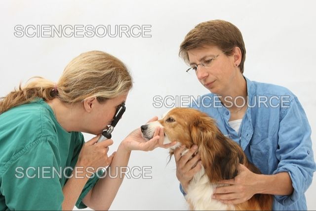
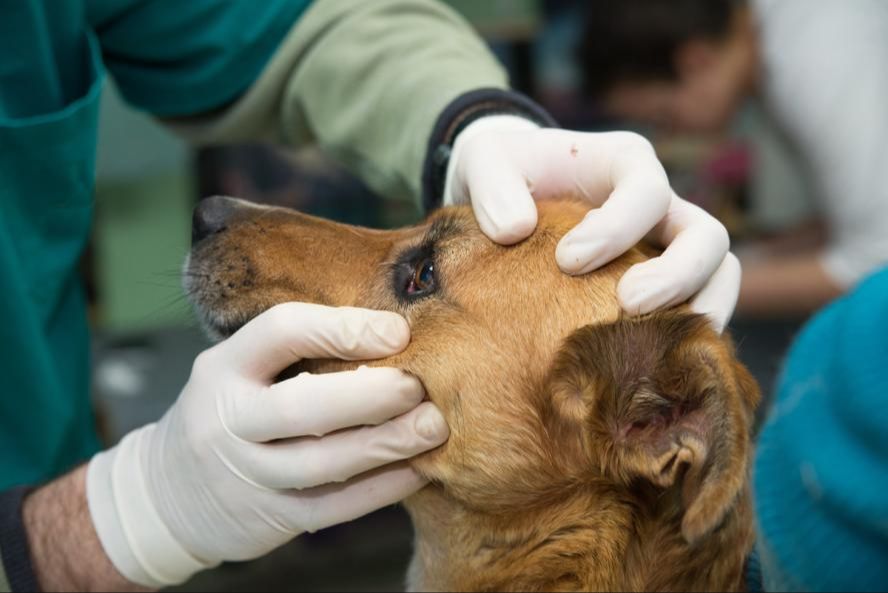
When to See the Vet
You should take your dog to the vet promptly if you notice any abnormal changes in the color of the third eyelid. Usually it is pink or has a pink tinge, so changes like it appearing red, dark red, purple, blue, or black warrant an exam. Additionally, if the third eyelid is visible when your dog’s eye is fully open, that is not normal and indicates an issue.
Other signs that indicate a vet visit is needed include swelling of the third eyelid, redness on the tissue around the eye, ocular discharge, or your dog squinting, pawing at its eye, or rubbing its face. Per Conditions of the Third Eyelid Factsheet, when owners notice any sudden change in the appearance of a dog’s eye, they should call their vet right away.
Since abnormal third eyelid changes may signal an eye condition or problem, it’s important to have your vet examine it promptly. Catching issues early can help prevent worsening symptoms and damage. Your vet can diagnose the cause and recommend proper treatment.
Diagnosing the Cause
Determining the underlying cause of third eyelid abnormalities typically involves a thorough medical evaluation by a veterinarian. This includes:
-
Medical history – The vet will ask about your dog’s health, any recent illnesses or injuries, medications, and potential exposure to irritants or allergens.
-
Physical exam – The vet will examine your dog’s eyes closely, looking for abnormalities of the third eyelid as well as the eyeball, eyelids, and surrounding structures.
-
Fluorescein stain test – This involves applying an orange dye to the eye that glows green under a special light. It can reveal ulcers, scratches or other damage to the cornea.
-
Biopsy – A small sample of the third eyelid may be taken and examined under a microscope. This can diagnose certain tumors, immune disorders, and other conditions.
-
Bloodwork – Blood tests can uncover issues like infections, autoimmune diseases, diabetes, and more.
Once a diagnosis is made, the vet will recommend appropriate treatment options tailored to the specific cause.
Treatment Options
The main treatment options for abnormalities of the third eyelid in dogs include:
- Medicated eye drops or ointments – Antibiotic and/or anti-inflammatory eye drops or ointment will be prescribed for 7-10 days to treat any infections or inflammation. Common options include neomycin, polymyxin B, bacitracin, and dexamethasone.
- Oral medications – Oral antibiotics and anti-inflammatories will usually be prescribed for 5-10 days to treat underlying causes and reduce swelling. Common oral options include amoxicillin, cephalexin, prednisone, and pain medications.
- Surgery – If the condition is caused by tumors or deep ulcers on the third eyelid, surgery may be required to remove damaged tissue. This can prevent discomfort and further issues.
The veterinarian will determine the right course of treatment based on the underlying cause and severity. Mild issues can often be treated with medicated drops, while chronic or tumorous changes may require surgery for a permanent fix. Follow up exams are crucial to ensure treatment is working.
Sources:
https://vetspecialists.co.uk/fact-sheets-post/conditions-of-the-third-eyelid/
https://www.ndsr.co.uk/information-sheets/cherry-eye-in-dogs-third-eyelid-gland-prolapse/
Home Care and Recovery
Proper aftercare at home is important for your dog’s recovery when being treated for third eyelid problems. Here are some tips:
Apply any prescribed eye drops or ointment as directed by your veterinarian. It is crucial to follow the medication schedule and dosage recommendations precisely. This helps treat the condition effectively and prevent recurrence (source).
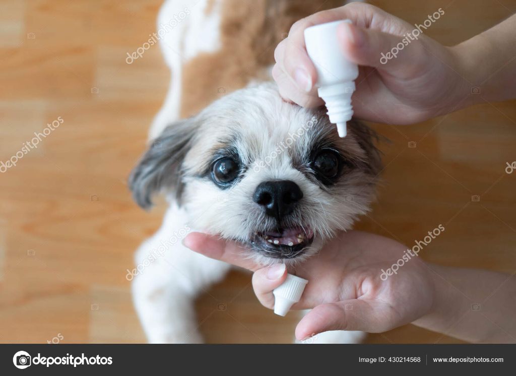
You may need to put an Elizabethan collar on your dog to prevent them from rubbing their eyes and dislodging medication. The collar allows the eyes time to heal without irritation (source).
Monitor your dog’s eyes closely over the recovery period. Look for any worsening of symptoms or new abnormalities. Contact your vet if the condition seems to deteriorate. Ongoing observation ensures proper healing.
Follow up with your veterinarian as recommended for rechecks and continued treatment if needed. Closely adhering to recovery guidelines gives your dog the best chance of overcoming third eyelid issues.
Prevention
There are some steps you can take to help prevent third eyelid issues in your dog:
- Avoid trauma and irritants – Be careful around your dog’s eyes and avoid exposing them to things like dirt, debris, chemicals, etc. that could cause irritation or injury (source).
- Monitor eye health at home – Check your dog’s eyes regularly for any abnormalities like redness, discharge, or swelling. Look to see if the third eyelid is protruding more than usual (source).
- Annual vet exams to catch issues early – Have your veterinarian examine your dog’s eyes closely each year and bring up any concerns you notice at home. Early treatment can help prevent more serious issues (source).
Staying attentive to your dog’s eye health and getting veterinary care when needed can help reduce the risks of third eyelid abnormalities.