Introduction
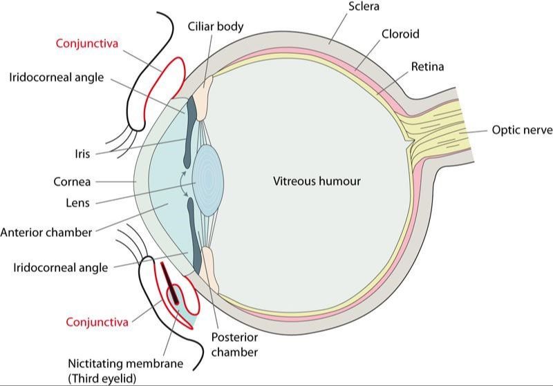
The third eyelid, also known as the nictitating membrane, is an extra eyelid found in many animals including dogs. It is located in the inner corner of the eye. The purpose of the third eyelid in dogs is to help protect, lubricate, and clean the eye.
The third eyelid acts as an extra barrier to protect the eye from irritants such as dust, dirt, and debris. It also helps spread tears and lubricate the surface of the eye, preventing it from drying out. When dogs blink, the third eyelid sweeps across the eye surface, helping to remove excess mucus, debris, and foreign bodies.
Overall, the third eyelid plays an important role in maintaining eye health and providing additional protection and moisture for the sensitive eye structures in dogs.
Anatomy
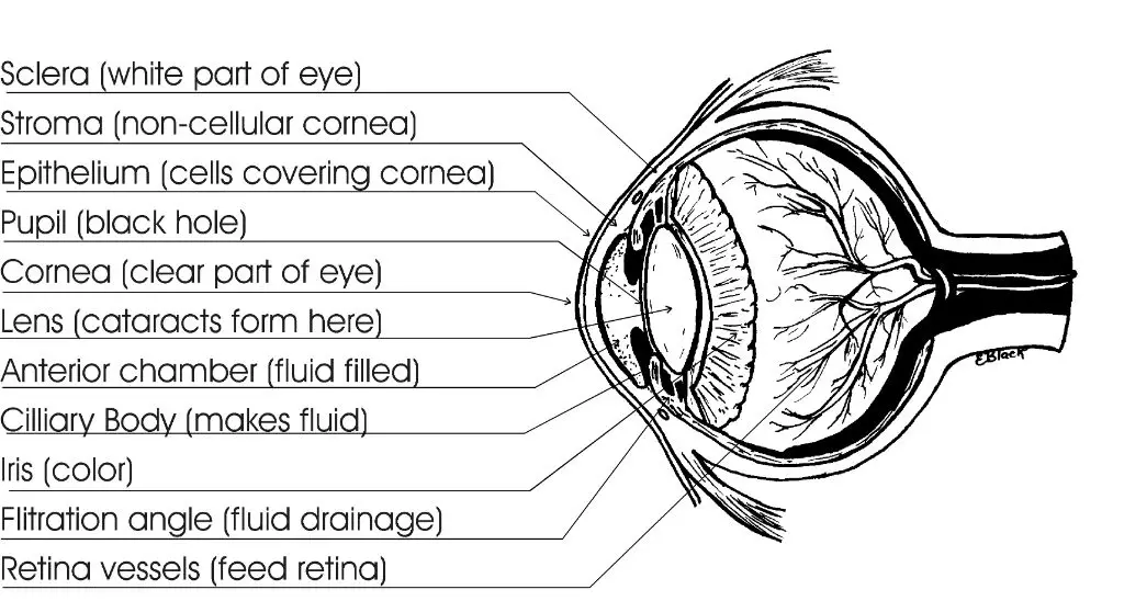
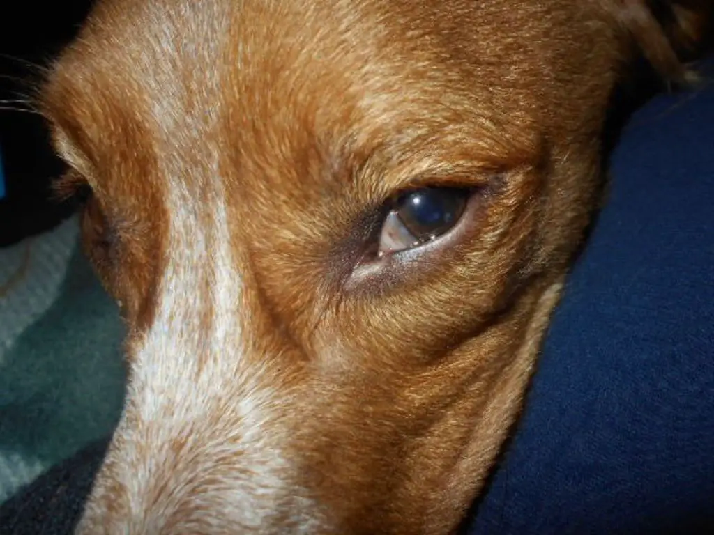
The technical name for the third eyelid is the nictitating membrane or palpebra tertia in dogs. It is located on the inner corner of a dog’s eye, underneath the lower eyelid. The third eyelid appears as a pink tissue that can extend across the eye.
According to an article on All About Vision, “In dogs, the nictitating membrane is located under the lower eyelid on the inner corner of the eye. Also called the haw or inner eyelid, a dog’s third eyelid functions to lubricate and protect the surface of the eye.” (Source).
Function
Dogs have a third eyelid located in the inner corner of their eye that serves several important functions. Its main role is for protection. The third eyelid protects the dog’s eye by acting as an additional barrier against irritants like dust, wind, and debris that could potentially scratch or damage the surface of the eye ([1]).
The third eyelid also helps keep the eye moist and lubricated by spreading tears across the surface of the eye. Tears are produced in the lacrimal gland above the eyeball and drain through small ducts into the conjunctival sac. As the third eyelid moves back and forth horizontally across the eye, it helps spread the tear film evenly over the cornea ([2]). This constant lubrication is essential in keeping the surface of the eye smooth and clear.
Finally, the third eyelid plays a role in tear drainage. There are small openings at the base of the third eyelid that allow excess tears to drain down through the nasolacrimal ducts and into the nasal passages. Without the third eyelid aiding this drainage, tears could potentially overflow from the eye ([1]).
Innervation
The main nerve supplying the third eyelid in dogs is the mandibular branch of the trigeminal nerve (https://veteriankey.com/third-eyelid-2/). This branch provides parasympathetic innervation to the membrane and gland. The parasympathetic fibers originate in the pterygopalatine ganglion and travel along the zygomatic nerve and zygomaticotemporal nerve to reach the third eyelid (https://www.sciencedirect.com/topics/veterinary-science-and-veterinary-medicine/nictitating-membrane). Stimulation of these nerves causes the third eyelid to protrude out and spread tears across the eye.
Sympathetic innervation of the third eyelid comes from the superior cervical ganglion. These nerves control retraction of the membrane. Damage to the sympathetic supply can cause Horner’s syndrome, leading to prolapse of the third eyelid.
Branches
The nerve to the third eyelid in dogs is part of the sympathetic nervous system, known as the ocular sympathetic nerve pathway. The ocular sympathetic nerve originates in the hypothalamus and descends through the brainstem and cervical spinal cord.
The nerve then follows three main branches (Source):
- The sympathetic trunk, which runs alongside the vertebral column
- The carotid nerve, which runs alongside the carotid artery in the neck
- The cavernous nerve, which enters the orbit and innervates the eye
Within the orbit, the postganglionic sympathetic fibers from the cavernous nerve supply the smooth muscle of the third eyelid, causing retraction when stimulated (Source). Damage or interruption anywhere along this pathway can result in Horner’s syndrome, characterized by protrusion of the third eyelid.
Control
The third eyelid in dogs is controlled by the retractor bulbi muscle. This muscle originates from the back of the eye socket and attaches to the third eyelid, allowing it to retract and protrude the membrane over the eye (Source).
When the retractor bulbi muscle contracts, it pulls the third eyelid towards the back of the eye socket, exposing the cornea. When the muscle relaxes, the third eyelid moves forward to cover part of the eye. This helps protect and lubricate the cornea.
The retractor bulbi muscle moves the third eyelid reflexively in response to stimuli that may damage the eye. For example, the membrane shoots forward when the eye is irritated or if the dog is suddenly startled. Conscious control over the third eyelid is limited in dogs.
Disorders
The third eyelid can be affected by several disorders that impact its normal function. Some common problems include:
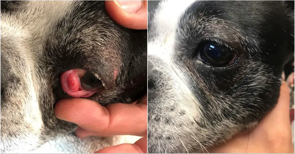
- Cherry eye – a prolapse of the third eyelid gland, causing the gland to swell and bulge out as a red mass in the corner of the eye.
- Entropion – the eyelid rolls inward, causing the eyelashes to rub against the eye.
- Ectropion – the eyelid droops or rolls outward.
- Conjunctivitis – inflammation of the conjunctiva, the pink tissue lining the eyelids and third eyelid.
- Scrolling – abnormal curling of the edge of the third eyelid.
- Horner’s syndrome – a neurological disorder that affects nerves controlling the eyelids.
- Cancer – third eyelid tumors are possible but rare.
These conditions can cause redness, swelling, discharge, squinting, and other signs of irritation or discomfort. Proper examination and treatment by a veterinarian is recommended if the third eyelid appears abnormal.
Examination
Veterinarians routinely examine a dog’s third eyelid as part of a full physical exam. They will look for any abnormalities in the appearance, position, or movement of the third eyelid that could indicate an underlying health issue.
To examine the third eyelid, the vet will gently pull down on the lower eyelid to expose the third eyelid. They will look to see if the third eyelid is protruding over the eye more than normal, or if it looks red, inflamed or thickened. The vet will also check if the third eyelid moves properly when touched. Sluggish or impaired movement could signal a problem with the nerves or muscles controlling the third eyelid.
Vets may also press on the eye to see if that causes the third eyelid to protrude more. Excessive protrusion when touching around the eye could indicate irritation or discomfort. Fluorescein dye may be applied to check for ulcers on the third eyelid or other parts of the eye if an issue is suspected.
According to the veterinary eye care experts at Washington State University, checking the third eyelid is an important part of monitoring eye health and detecting problems early on [1]. Catching issues with the third eyelid promptly can help prevent more serious eye damage and loss of vision.
Treatment
There are a few options for treating disorders of the third eyelid in dogs:
Surgery is often recommended to reposition the gland if it has prolapsed and is causing a “cherry eye.” According to VCA Animal Hospitals, “Treatment involves surgical replacement of the third eyelid gland. It is essential to treat the condition as soon as possible to prevent secondary inflammation and thickening of the gland.”
The NDSR notes that surgery is the preferred treatment for third eyelid gland prolapse: “The recommended treatment is surgery to replace the gland to its normal position at the base of the third eyelid where it originated.”
Medications may also be prescribed, such as antibiotic or anti-inflammatory eye drops/ointment for 7-10 days along with oral medication for 5-10 days according to Vetspecialists.co.uk.
In some cases, the underlying cause may need to be addressed, such as treating allergies if they are causing inflammation of the third eyelid.
Summary
The third eyelid, also known as the nictitating membrane or haw, plays an important role in maintaining eye health and providing additional protection in dogs. This inner eyelid is primarily controlled by the sympathetic nervous system and innervated by the mandibular branch of the trigeminal nerve (cranial nerve V).
Specifically, parasympathetic fibers originate from the pterygopalatine ganglion and travel to the third eyelid via the zygomatic nerve and a branch of the maxillary nerve. Sympathetic innervation stems from the superior cervical ganglion and runs along the internal carotid artery. Together, these nerves allow for rapid protraction and retraction of the third eyelid.
Problems with third eyelid innervation or function can lead to prolapsed gland of the third eyelid and other disorders. Careful examination and imaging can help diagnose underlying issues. Treatment focuses on lubrication, anti-inflammatories, and surgery in severe cases. Overall, the intricate innervation of the canine third eyelid allows this unique structure to help protect the eye.
