Introduction
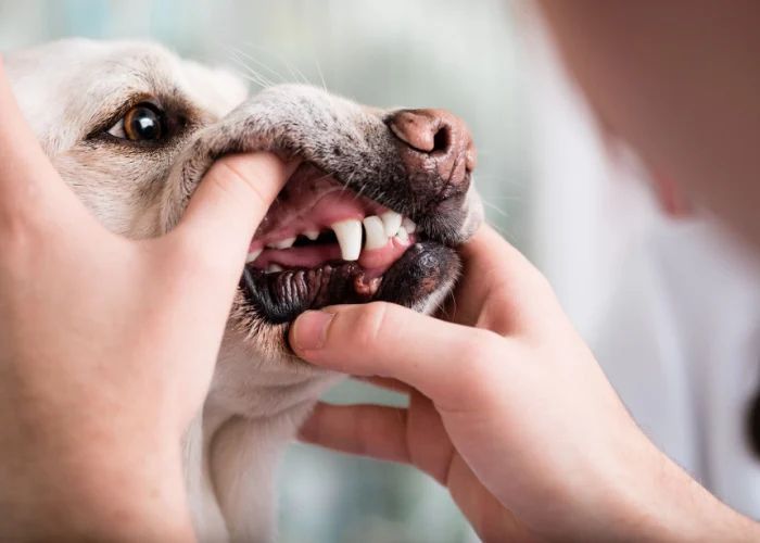
Proper dental care is an important part of every dog’s overall health and wellbeing. Like humans, dogs can develop tartar buildup, gum disease, cavities, and other oral health issues over time. In some cases, severely damaged or infected teeth may need to be extracted by a veterinarian.
The process of extracting a tooth is usually quick but requires anesthesia and follow-up care. Not all teeth are equally easy to remove – some extractions are straightforward while others carry greater risks and potential complications.
One of the most challenging teeth to extract in dogs is the upper fourth premolar. This molar tooth has an extensive root system and is positioned at the back of the mouth, making access difficult. Removing it takes skill and care to avoid fracturing the jawbone or leaving root fragments behind.
In this article, we’ll look at why the upper fourth premolar is often considered the most troublesome tooth for extractions in dogs. We’ll also go over the extraction procedure, aftercare, costs, and risks involved.
Anatomy of the Dog Mouth
Dogs have a total of 42 teeth, depending on the breed. These consist of incisors, canines, premolars and molars located in the upper jaw and lower jaw.
Incisors are the small teeth at the front of the mouth used for biting and tearing food. Dogs have 6 upper incisors and 6 lower incisors.
Canines, also known as fangs, are located behind the incisors and are used for holding and tearing prey. Dogs have 4 canine teeth – 2 in the upper jaw and 2 in the lower jaw.
Premolars are located behind the canines and have sharper edges useful for grinding food. Dogs have 8 upper premolars and 8 lower premolars.
Molars are the large teeth at the back of the mouth used for chewing and crushing food. Dogs have 2 upper molars and 3 lower molars.
Knowing the anatomy and purpose of the different types of teeth in a dog’s mouth provides context for understanding extraction challenges.
Most Difficult Tooth to Extract
The most difficult tooth to extract in dogs is generally agreed to be the maxillary fourth premolar (upper fourth premolar). This tooth is located in the upper back portion of the mouth. There are several reasons why the maxillary fourth premolar presents unique challenges compared to other teeth:
The root of this tooth is positioned at an angle which requires the veterinarian to maneuver around other teeth and bone structures. The angled root combined with its sturdy anchor in the bone makes the tooth more resistant to extraction forces.
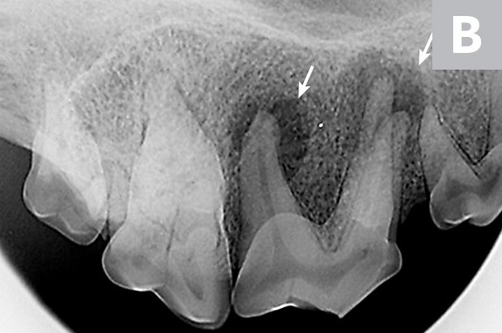
Additionally, the maxillary fourth premolar often has multiple roots. The presence of multiple roots complicates the extraction process and requires more care to fully remove the tooth and all root material. Fragments of root left behind can cause ongoing dental issues.
The shape, angle, and root anatomy of the maxillary fourth premolar makes it the most technically challenging tooth for veterinarians to fully and safely extract from dogs. Proper extraction of this tooth requires precision, skill, and the right tools to elevate and section the tooth for complete removal.
Why the Fourth Premolar is Difficult
The canine fourth premolar is one of the hardest to extract teeth in dogs due to several factors:
The roots of the fourth premolar are curved, which makes them difficult to extract without breaking. The tooth’s roots are firmly anchored in the jaw bone, so extracting them requires substantial force. This curvature means extra care must be taken when loosening and removing the tooth.
The fourth premolar is also positioned at an awkward angle in the back of the mouth behind the large carnassial tooth. The angle and location make it hard for veterinarians to get proper leverage and access to the tooth during extraction.
Importantly, the positioning of the fourth premolar and its curved roots means there is a higher risk of jaw fracture during extraction. Veterinarians have to take care to avoid applying too much pressure and potentially cracking the mandible bone. Fracturing the jaw would lead to additional complications and a more involved recovery.
In summary, the curved roots, angled positioning, and fracture risk make extracting the canine fourth premolar one of the most difficult and delicate dental procedures veterinarians perform.
Extraction Process
The extraction process for the difficult fourth premolar tooth involves sedation and surgical extraction. Here are the main steps:
Sedation – The dog will be given a sedative by injection or inhaled gas to make them relaxed and sleepy for the procedure. This ensures the dog stays still and comfortable.
Anesthetic – A local anesthetic is given by injection near the extraction site to numb the area so the dog feels no pain.
Incision – The vet will make a small incision in the gum around the tooth to access it.
Tooth removal – The tooth is loosened from the socket, often by rocking it back and forth. The vet may need to cut the ligaments attaching it with a scalpel.
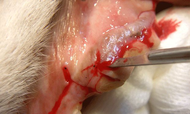
Sutures – The gum is sutured closed with stitches that will dissolve over time.
Recovery – The dog is monitored until fully awake from sedation. Pain medication may be prescribed for a few days.
Aftercare
After a dog has had the fourth premolar tooth extracted, proper aftercare is crucial for a smooth recovery. Owners will need to closely monitor their dog and follow all instructions from the veterinarian. Here are some of the main aspects of aftercare following fourth premolar extraction surgery:
Pain Management
The vet will prescribe pain medication, typically an NSAID like Rimadyl, to keep the dog comfortable after surgery. It’s important to give this medication exactly as directed. Restrict activity and don’t let the dog play roughly while recovering. Use a soft recovery collar to prevent irritating the surgical site.
Antibiotics
Antibiotics may be prescribed to prevent infection after extraction. Be sure to finish the entire course as directed, usually 7-10 days. Call the vet if you notice signs of infection like swelling, discharge or fever.
Soft Food Diet
After losing a premolar tooth, dogs should eat soft, moist food for 2 weeks or so. Canned food or kibble soaked in warm water or broth works well. Avoid anything hard or crunchy that could damage the surgical site while it’s healing. Gradually reintroduce dry kibble once healing is complete.
Cost of Fourth Premolar Extraction
The cost of extracting the fourth premolar tooth in dogs can vary depending on your location and the veterinarian performing the procedure. However, on average, owners can expect to pay between $200-$800 to have this challenging tooth removed.
Some of the factors that influence the cost include:
- Your geographical location – Urban clinics will charge more than rural ones
- The age and health of your dog – More complex extractions cost more
- Whether your dog is undergoing other procedures – Costs may be bundled
- The veterinarian’s experience and specialization
- Whether general anesthesia is required
- Aftercare medications and instructions
While the upper range may seem high, extracting the fourth premolar is complex and requires the expertise of a veterinary dental specialist. This delicate procedure often takes 1-2 hours to complete. Ultimately, paying more for an experienced veterinarian can reduce complications and the need for additional procedures later on.
Risks and Complications
Extracting the fourth premolar tooth in dogs does carry some risks that pet owners should be aware of before the procedure. The main potential complications include:
Nerve Damage
There are many nerves that run through the jaw area that allow sensation and function of the face. During extraction, these nerves can potentially be damaged. This may lead to numbness or altered sensation in the lips, chin or tongue after surgery. Nerve damage can sometimes be temporary as the nerves heal, but in some cases it can be permanent.
Jaw Fracture

The mandible (lower jaw) contains a thin section of bone where the premolar roots are located. Applying too much force during extraction can lead to a fracture in this area. Jaw fractures often require surgical repair and several weeks of healing time.
Infection
Anytime surgery is performed inside the mouth, there is potential for bacteria to enter the wound and cause an infection. Signs of infection include swelling, redness, foul odor from the mouth and discharge. Infections require antibiotic treatment and can delay healing. Proper oral care after surgery is important to prevent infection.
Alternatives to Extraction
Before deciding to extract a tooth, especially one as difficult as the fourth premolar, it’s important to consider alternatives that may allow the tooth to be saved. Two of the main alternatives are root canal therapy and crown placement.
A root canal involves removing the infected pulp from inside the tooth and sealing it off. This eliminates the infection and allows the tooth to heal and remain in place. Root canals can be done on any tooth, including premolars. Success rates for dog root canals are over 90%.
After a root canal, a crown is placed over the remaining tooth to protect it from fracturing. The crown covers the tooth above the gum line and acts as an artificial covering. Crowns can prevent extraction in cases where the tooth is damaged but the root is still healthy.
For fourth premolars with deep decay, infections, or fractures, a root canal and crown may spare the dog from losing the tooth entirely. This should be discussed with your veterinarian as an alternative to extraction in appropriate cases.
When Extraction is Necessary
There are a few instances when extracting the fourth premolar becomes absolutely necessary in dogs:
- Severe Periodontal Disease – If the tooth is severely affected by advanced periodontal disease that has destroyed the periodontal ligament and alveolar bone, extraction is required. The disease will continue to spread if the tooth is not removed.
- Impacted/Damaged Tooth – An impacted or damaged fourth premolar that cannot be saved through restorative dental work needs to be extracted. Leaving the tooth in place will likely cause pain, dental problems and infection.
In these cases, the benefits of extraction outweigh the risks. Though removing the fourth premolar is complicated, extraction helps eliminate sources of infection and discomfort. It is an important treatment option for certain dental issues in dogs.