Introduction
An abdominal ultrasound, also referred to as a sonogram, is an imaging technique that uses high-frequency sound waves to create images of the internal organs and structures in a dog’s abdomen. The purpose of this article is to explain why a veterinarian may recommend an abdominal ultrasound for a dog and what conditions it can help diagnose.
Evaluating Abdominal Pain or Discomfort

An abdominal ultrasound can be a very useful tool for evaluating a dog experiencing abdominal pain or discomfort. The ultrasound allows the veterinarian to get a detailed look inside the dog’s abdomen to try to pinpoint the underlying cause.
Some potential causes of abdominal pain that an ultrasound can help diagnose include:
- Pancreatitis – inflammation of the pancreas
- Intestinal foreign body – an ingested object lodged in the intestines
- Intestinal obstruction – a blockage or twist in the intestines
- Gastrointestinal ulceration – ulcers in the stomach or intestines
- Inflammatory bowel disease – chronic intestinal inflammation
- Abdominal mass or tumor – such as cancer or benign growths
- Abdominal fluid accumulation – blood, pus, bile or other fluids
- Liver or spleen abnormalities – infections, masses, cirrhosis
- Bladder stones or obstruction
- Kidney issues – infection, stones, masses, failure
The ultrasound allows the veterinarian to look for abnormalities in the organs, view any free fluid in the abdomen, and identify potential sources of pain that may require further testing or treatment. It is an invaluable tool for determining the underlying cause in a dog with abdominal discomfort.
Assessing Organ Health
One of the main uses for abdominal ultrasounds in dogs is to evaluate the health and appearance of internal organs like the liver, kidneys, and spleen. The ultrasound waves can penetrate the abdominal tissues and muscles to provide clear images of these organs without any invasive surgery.
Looking at the size, shape, and texture of the organs helps veterinarians determine if there are any abnormalities present. For example, an enlarged spleen may indicate splenomegaly or extramedullary hematopoiesis. The kidneys may appear abnormal in size or shape if there is kidney disease, hydronephrosis or even cancer present. Veterinarians can also use Doppler ultrasound to assess blood flow within the organs.
Visualizing the internal organs is crucial for identifying potential sources of illnesses and ensuring the organs are functioning properly. It allows for earlier diagnosis and treatment of many conditions compared to just a physical exam alone. According to Sunstone Vets, ultrasound has nearly 100% sensitivity for detecting anatomical changes and abnormalities in abdominal organs. [1]
Overall, the detailed organ evaluation possible through abdominal ultrasound exams makes them a very useful diagnostic tool for veterinarians to analyze a dog’s health and determine next steps for care.
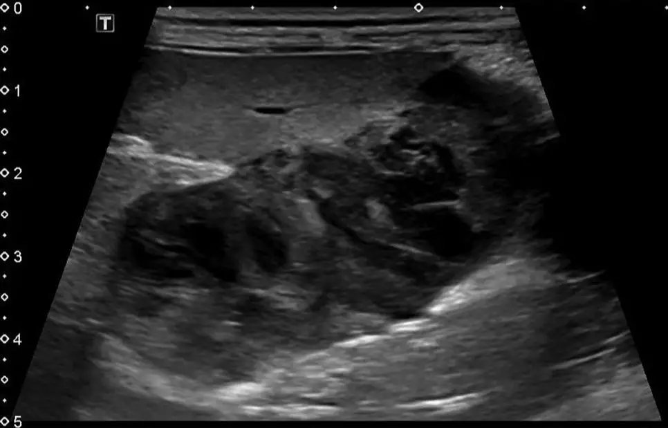
Identifying Masses or Tumors
Ultrasound is one of the most accurate ways to detect masses or tumors in a dog’s abdomen, even at an early stage. The high-frequency sound waves used in ultrasound can provide detailed images of organs and tissues, allowing a veterinarian to identify any abnormal growths or structures. Ultrasound has been shown to detect masses as small as 2-3mm in diameter.
One key advantage of ultrasound for detecting abdominal masses is that it allows the veterinarian to see the exact size, shape, location and characteristics of a mass. This helps determine if it is a benign growth or potentially cancerous tumor. Ultrasound can identify features of malignant tumors like irregular margins, heterogeneity, and vascularity.
Compared to just feeling for masses during a physical exam, ultrasound provides far more detail. It also does not require sedation, unlike more invasive tests like exploratory surgery. This makes it a convenient first-line tool for mass detection. According to one study, gastric masses were detected in 91% of cases using abdominal ultrasound in dogs presenting with vomiting (source).
Ultrasound is considered one of the best ways to monitor tumors and check for recurrence after treatment. It allows veterinarians to closely track the size and features of any persistent masses. This helps guide treatment decisions and provide prognoses for canine patients.
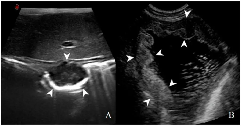
Checking for Free Fluid
One of the most common reasons for performing an abdominal ultrasound in dogs is to check for free fluid, which may indicate bleeding or infection in the abdomen and abdominal cavity (the peritoneal space). Ultrasound is very sensitive at detecting even small amounts of free fluid, appearing as black or anechoic regions on the ultrasound image (source). The presence of free fluid often prompts further tests like abdominocentesis to analyze the fluid. One study found ultrasound had 97% sensitivity and 83% specificity for detecting free abdominal fluid compared to abdominocentesis as the gold standard (source).
Free fluid may accumulate from leakage of urine, blood, bile, intestinal contents, ascites, or other sources. Determining the cause of the fluid buildup is important. Hemorrhage, trauma, neoplasia, coagulopathy, infection, inflammation, hepatic failure, cardiac failure, or other conditions could result in free fluid. Ultrasound allows veterinarians to non-invasively visualize and monitor free fluid over time. This aids in diagnosis, gauging the severity, and tracking the treatment response (source).
Monitoring Pregnancy
An abdominal ultrasound is an important tool for monitoring pregnancy and fetal health in dogs. Ultrasound allows veterinarians to confirm pregnancy as early as 3-4 weeks after breeding https://www.eastcentralvet.com/canine-pregnancy.pml. While not as accurate for counting puppies, ultrasound provides an overall assessment of fetal size and development.
Ultrasound is used to check for pregnancy signs like fetal heartbeats and fetal movement. It can detect potential issues like uterine inflammation, fetal abnormalities, or resorbing fetuses. Sequential ultrasounds allow vets to monitor progress and growth. Ultrasound is more effective for monitoring early pregnancy than x-rays which are better later in gestation https://www.bcfultrasound.com/canine-pregnancy-part-1-radiography-vs-ultrasound/.
Overall, ultrasound is a safe, non-invasive way for vets to confirm pregnancy in dogs and monitor fetal development and wellbeing throughout gestation.
Guiding Medical Procedures
Ultrasound is often used to guide minimally invasive procedures like fine needle aspirates, biopsies, and drainages in dogs (Ultrasound-Guided Procedures). The real-time imaging allows veterinarians to identify the exact location for needle insertion and monitor the needle’s path to the target area. This improves accuracy and reduces complications. Some common procedures guided by ultrasound include:
- Fine needle aspiration of organs or masses to collect samples for cytology
- Biopsies of organs like the liver, kidneys, and spleen
- Drainage of abscesses or fluid accumulations
- Placement of feeding tubes
Ultrasound guidance ensures proper needle positioning, avoiding damage to surrounding tissues and vessels. It also allows screening during the procedure to identify any complications. The visual feedback enables real-time adjustments to improve the safety and success of needle-based procedures in dogs.
Follow-up Exams
Abdominal ultrasound can be a useful tool in monitoring certain conditions over time and evaluating a patient’s response to treatment. Some common reasons for follow-up abdominal ultrasound exams in dogs include:
Monitoring cancers or tumors – Ultrasound allows veterinarians to periodically check the size and spread of tumors or masses to determine if they are responding to chemotherapy or radiation treatments (Webb 2012).
Checking pregnancy – Ultrasounds every 2-3 weeks can track fetal growth and development, and screen for potential complications during the pregnancy (Hampstead Animal Hospital).
Assessing organ health – Conditions affecting the liver, kidneys, spleen, bladder and other organs can be periodically evaluated with ultrasound to monitor disease progression or improvement.
Monitoring free abdominal fluid – Free fluid in the abdomen may indicate bleeding, infection, cancer, liver disease, and other conditions. Follow-up ultrasounds can track changes in free fluid over time.
Overall, abdominal ultrasound allows for regular re-evaluation of a patient’s health and specific conditions. This helps veterinarians determine the effectiveness of treatments and make informed decisions about ongoing care.
Limitations of Abdominal Ultrasound
While abdominal ultrasound is a versatile and useful diagnostic tool, there are some limitations to be aware of (VCA Hospitals):
Ultrasound waves do not penetrate gas or bone well, so imaging of organs obscured by the ribs or those containing gas, like the intestines, may be limited. Obese animals can also be difficult to image properly due to thicker layers of fat tissue blocking the ultrasound waves.
Ultrasound is very operator dependent and requires an experienced ultrasonographer to obtain quality images and make accurate interpretations. Results can vary between different operators.
Compared to other imaging modalities like CT or MRI, ultrasound provides less fine detail on organ structure and anatomy. Small tumors or masses may not be detectable on ultrasound. It is also not as effective at evaluating lymph nodes.
While ultrasound guided needle biopsies are possible, the sampling area is relatively small. It may miss sampling the most diagnostically useful area of a mass.
As useful as abdominal ultrasound is for pregnancy monitoring, it cannot reliably confirm pregnancy until approximately day 25-30 after breeding. Earlier than that, it may miss a pregnancy if the fetuses are too small to visualize.
In summary, ultrasound has limits when evaluating gas-filled viscera, small structures, lymph nodes, and early pregnancies. Operator skill is also a factor. Other imaging techniques may be preferable depending on the clinical question.
Conclusion
In summary, there are a variety of reasons why a veterinarian may recommend an abdominal ultrasound for a dog. This imaging technique allows the vet to evaluate organs, identify masses, check for fluid buildup, monitor pregnancy, guide procedures, and conduct follow-up exams. While abdominal ultrasounds have limitations and cannot detect all conditions, they provide a non-invasive way to gain valuable information about a dog’s abdominal health and identify potential problems requiring further diagnosis or treatment.
Some of the main reasons for performing abdominal ultrasounds on dogs include: assessing gastrointestinal issues like vomiting or diarrhea, checking major organs like the liver or kidneys, looking for tumors or cancer, diagnosing pregnancy and monitoring fetal health, guiding needle biopsies, guiding surgeries, and tracking post-procedure healing. This safe, relatively low-cost test allows vets to look inside a dog’s abdomen and provides key insights to determine next steps in care.
In the end, abdominal ultrasounds are a useful diagnostic tool in a veterinarian’s toolkit for evaluating a dog’s abdominal symptoms and guiding treatment decisions. While not a stand-alone panacea, ultrasounds provide vets and pet owners with valuable information to provide optimal care for canine patients.